BiP (タンパク質)
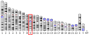
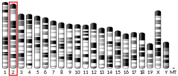
BiP(binding immunoglobulin protein)は、ヒトでは HSPA5遺伝子によってコードされるタンパク質である。GRP-78、HSPA5(heat shock 70 kDa protein 5)、Byun1としても知られる[5][6]。
BiPは、小胞体の内腔に位置するHsp70ファミリーの分子シャペロンである。小胞体へ移行してきた新生タンパク質に結合し、それらをその後のフォールティングやオリゴマー化が可能な状態に維持する。また、BiPは小胞体の移行装置の必須の構成要素でもあり、異常タンパク質のプロテアソーム分解へ向けた小胞体への逆行性輸送に役割を果たす。BiPはすべての生育条件で豊富にみられるタンパク質であるが、フォールディングしていないポリペプチドが小胞体に蓄積する条件下では合成が顕著に誘導される。
構造
[編集]BiPは、ヌクレオチド結合ドメイン(NBD)と基質結合ドメイン(SBD)という2つの機能的ドメインを含んでいる。NBDはATPを結合して加水分解し、SBDはポリペプチドを結合する[7]。
NBDは2つの大きな球状サブドメイン(I、II)から構成され、さらにそのそれぞれが2つの小さなサブドメイン(A、B)へと分割される。サブドメイン間には溝があり、そこへヌクレオチド、1つのMg2+イオン、2つのK+イオンが結合して4つのドメイン(IA、IB、IIA、IIB)すべてが連結される[8][9][10]。SBDは、SBDβとSBDαという2つのサブドメインへと分割される。SBDβは基質タンパク質またはペプチドの結合ポケットとして機能し、SBDαは結合ポケットを覆うαヘリックスからなる蓋として機能する[11][12][13]。ドメイン間リンカーはNBDとSBDを連結し、NBD-SBD相互作用面の形成を促進する[7]。
機構
[編集]BiPの活性は、アロステリックなATPアーゼサイクルによって調節されている。ATPがNBDに結合するとSBDαの蓋が開き、SBDは基質との親和性が低いコンフォメーションとなる。ATPの加水分解に伴ってADPがNBDに結合し、蓋が結合した基質の上に閉じる。これによって基質は解離速度が低下し高い親和性での結合を行い、基質の尚早なフォールディングや凝集が防がれる。ADPがATPへの交換されるとSBDαの蓋が開いて基質が放出され、基質は自由にフォールディングを行うようになる[14][15][16]。ATPアーゼサイクルは、プロテインジスルフィドイソメラーゼ[17]やコシャペロン[18]によって相乗的に加速される。
機能
[編集]細胞がグルコース飢餓にさらされると、グルコース調節タンパク質(glucose-regulated protein、GRP)と呼ばれるいくつかのタンパク質の合成が顕著に上昇する。GRP78(HSPA5)はBiPとも呼ばれ、Hsp70ファミリーのメンバーであり小胞体でのタンパク質のフォールディングや組み立てに関与する[6]。BiPのレベルは、小胞体内の分泌タンパク質(IgGなど)の量と強く相関している[19]。
BiPによる基質の解離と結合は、新生タンパク質のフォールディングや組み立て、誤ってフォールディングしたタンパク質の凝集の防止、分泌タンパク質の移行、小胞体ストレス応答(unfolded protein response、UPR)の開始など、小胞体での多様な機能を促進する。
タンパク質のフォールディングと保持
[編集]BiPは能動的に基質をフォールディングする(フォールダーゼ)、または単に結合して基質がフォールディングや凝集するのを防ぐ(ホルダーゼ)。フォールダーゼとして機能するには、完全なATPアーゼ活性とペプチド結合活性が必要である。ATPアーゼ活性に欠陥のある温度感受性変異体(クラスI変異と呼ばれる)とペプチド結合活性に欠陥のある変異体(クラスII変異と呼ばれる)は、どちらも非許容温度下ではカルボキシペプチダーゼY(CPY)を正しくフォールディングすることができない[20]。
小胞体への移行
[編集]BiPは小胞体の分子シャペロンとして機能し、小胞体内腔や小胞体膜へのATP依存的なポリペプチドの取り込みに必要である。ATPアーゼ活性変異体は、多数のタンパク質(インベルターゼ、カルボキシペプチダーゼY、α-接合因子)の小胞体内腔への移行の妨げとなることが判明している[21][22][23]。
小胞体関連分解
[編集]BiPは小胞体関連分解(ERAD)にも役割を果たす。最もよく研究されているERADの基質は、常に誤ったフォールディングを行い、完全に小胞体へ移行しグリコシル化修飾を受けるCPY(CPY*)である。BiPはCPY*と接触する最初のシャペロンで、CPY*の分解に必要とされる[24]。BiPのATPアーゼ活性変異(アロステリック変異も含む)によって、CPY*の分解速度が大きく低下することが示されている[25][26]。
小胞体ストレス応答経路
[編集]BiPはUPRの標的であるとともに、UPR経路に必須の調節因子でもある[27][28]。小胞体ストレス下では、BiPは3つのシグナル伝達因子(IRE1、PERK、ATF6)から解離し、効率的にそれぞれのUPR経路を活性化する[29]。BiPはUPRの標的遺伝子の産物であり、UPR転写因子がBiPの遺伝子DNAのプロモーター領域のUPRエレメントに結合することでアップレギュレーションされる[30]。
相互作用
[編集]BiPのATPaseサイクルは、ADPの解離の際にATPの結合を促進するヌクレオチド交換因子と、ATPの加水分解を促進するJタンパク質の双方のコシャペロンによって促進される[18]。
BiPのシステインの保存性
[編集]BiPは真核生物の間で高度に保存されており、それには哺乳類も含まれる(表1)。また、ヒトではBiPはすべての組織で広く発現している[31]。ヒトのBiPには2つの高度に保存されたシステイン残基が存在する。これらのシステイン残基は酵母と哺乳類細胞の双方で翻訳後修飾を受けることが示されている[32][33][34]。酵母細胞では、N末端側のメチオニンは酸化ストレスによってスルフェニル化とグルタチオン化されることが示されている。どちらの修飾も、BiPのタンパク質凝集を防ぐ能力を向上させる[32][33]。マウスの細胞では、保存されたシステインのペアはGPX7(NPGPx)の活性化に伴ってジスルフィド結合を形成する。ジスルフィド結合はBiPの変性タンパク質への結合を向上させる[35]。
| 表1.哺乳類細胞におけるBiPの保存性 | |||||
| 一般名 | 学名 | BiPの保存性 | BiPのシステインの保存性 | システイン数 | |
| 霊長類 | ヒト | Homo sapiens | Yes | Yes | 2 |
| ニホンザル | Macaca fuscata | Yes | Yes | 2 | |
| ミドリザル | Chlorocebus sabaeus | Predicted* | Yes | 2 | |
| マーモセット | Callithrix jacchus | Yes | Yes | 2 | |
| 齧歯類 | マウス | Mus musculus | Yes | Yes | 2 |
| ラット | Rattus norvegicus | Yes | Yes | 3 | |
| モルモット | Cavia porcellus | Predicted | Yes | 3 | |
| ハダカデバネズミ | Heterocephalus glaber | Yes | Yes | 3 | |
| ウサギ | Oryctolagus cuniculus | Predicted | Yes | 2 | |
| ツパイ | Tupaia chinensis | Yes | Yes | 2 | |
| 有蹄類 | ウシ | Bos taurus | Yes | Yes | 2 |
| ミンククジラ | Balaenoptera acutorostrata scammoni | Yes | Yes | 2 | |
| ブタ | Sus scrofa | Predicted | Yes | 2 | |
| 食肉類 | イヌ | Canis familiaris | Predicted | Yes | 2 |
| ネコ | Felis silvestris | Yes | Yes | 3 | |
| フェレット | Mustela putorius furo | Predicted | Yes | 2 | |
| 有袋類 | オポッサム | Monodelphis domestica | Predicted | Yes | 2 |
| タスマニアデビル | Sarcophilus harrisii | Predicted | Yes | 2 | |
| *Predicted: NCBI Proteinによる配列予測 | |||||
臨床的重要性
[編集]自己免疫疾患
[編集]ストレスタンパク質や熱ショックタンパク質の多くと同様、BiPは細胞内部環境から細胞外空間へ放出された際に強力な免疫学的活性を有している[36]。特に、BiPは免疫ネットワークへ抗炎症シグナルと解除促進シグナルを送り、炎症の解消を助ける[37]。BiPの免疫活性における機構は完全には理解されていない。しかしながら、単球表面の受容体に結合して抗炎症サイトカインの分泌を誘導し、T細胞の活性化に関わる重要な分子をダウンレギュレーションするとともに、単球の樹状細胞への分化経路を調節することが示されている[38][39]。
BiP/GRP78の強力な免疫調節活性は、ヒトの関節リウマチに似たマウス疾患であるコラーゲン誘導関節炎などの自己免疫疾患の動物モデルで示されている[40]。BiPの予防的・治療的な非経口デリバリーは、炎症性関節炎の臨床的・組織学的徴候を緩和することが示されている[41]。
心血管疾患
[編集]BiPのアップレギュレーションは、小胞体ストレスによって誘導される心機能不全や拡張型心筋症と関係している[42][43]。またBiPは、ホモシステイン誘導性の小胞体ストレスの緩和、血管内皮細胞のアポトーシスの防止、コレステロール/トリグリセリドの生合成を担う遺伝子の活性化の阻害、そして組織因子の凝血促進活性の抑制によって、アテローム性動脈硬化の発症を抑えると提唱されている。これらはすべてアテローム斑の蓄積に寄与する過程である[44]。
プロテアソーム阻害剤など一部の抗がん剤は、心不全の合併症と関係している。新生ラットの心筋細胞では、BiPの過剰発現はプロテアソームの阻害によって誘導される心筋細胞の細胞死を減少させる[45]。
神経変性疾患
[編集]BiPは小胞体のシャペロンタンパク質であり、誤ってフォールディングしたタンパク質を修正することで小胞体ストレスによる神経細胞の細胞死を防止する[46][47]。さらに、BIXと名付けられたBiPを誘導する化学物質は脳虚血モデルマウスで脳梗塞を減少させる[48][49]。逆に、BiPのシャペロン機能の向上はアルツハイマー病と強く関係している[44][49]。
代謝性疾患
[編集]BiPのヘテロ接合性は、小胞体ストレス経路をアップレギュレーションし、高脂肪食による肥満、2型糖尿病、膵炎から保護すると提唱されている。また、BiPは脂肪組織における脂肪生成とグルコースの恒常性に必要である[50]。
感染症
[編集]原核生物のBiPのオルソログは、細菌のDNA複製に必須なRecAなどのタンパク質と相互作用することが判明している。したがって、これらの細菌のHsp70型シャペロンは抗生物質開発の有望な標的となる。特にBiPを抑制する抗がん剤OSU-03012によって、淋菌Neisseria gonorrhoeaeのスーパー耐性菌(superbug)株はいくつかの標準的治療で用いられる抗生物質に対し再感受性となる[49]。一方志賀毒素産生性大腸菌の病原性株は、宿主のBiPを阻害するためにAB5型トキシンを産生し、宿主細胞の生存を弱体化させる[44]。対照的にウイルスは、細胞表面のBiPを介して細胞に感染し、ウイルスタンパク質へのシャペロン活性のためにBiPの発現を促進し、小胞体ストレスによる細胞死応答を抑制するなど、複製の大部分を宿主のBiPに依存している[49][51]。
出典
[編集]- ^ a b c GRCh38: Ensembl release 89: ENSG00000044574 - Ensembl, May 2017
- ^ a b c GRCm38: Ensembl release 89: ENSMUSG00000026864 - Ensembl, May 2017
- ^ Human PubMed Reference:
- ^ Mouse PubMed Reference:
- ^ “Human gene encoding the 78,000-dalton glucose-regulated protein and its pseudogene: structure, conservation, and regulation”. DNA 7 (4): 275–86. (May 1988). doi:10.1089/dna.1988.7.275. PMID 2840249.
- ^ a b “Localization of the gene encoding human BiP/GRP78, the endoplasmic reticulum cognate of the HSP70 family, to chromosome 9q34”. Genomics 20 (2): 281–4. (Mar 1994). doi:10.1006/geno.1994.1166. PMID 8020977.
- ^ a b “Close and Allosteric Opening of the Polypeptide-Binding Site in a Human Hsp70 Chaperone BiP”. Structure 23 (12): 2191–203. (Dec 2015). doi:10.1016/j.str.2015.10.012. PMC 4680848. PMID 26655470.
- ^ “1H, 13C, and 15N backbone assignment and secondary structure of the receptor-binding domain of vascular endothelial growth factor”. Protein Science 6 (10): 2250–60. (Oct 1997). doi:10.1002/pro.5560061020. PMC 2143562. PMID 9336848.
- ^ “Hsp70 chaperones: cellular functions and molecular mechanism”. Cellular and Molecular Life Sciences 62 (6): 670–84. (Mar 2005). doi:10.1007/s00018-004-4464-6. PMC 2773841. PMID 15770419.
- ^ “Crystal structures of the ATPase domains of four human Hsp70 isoforms: HSPA1L/Hsp70-hom, HSPA2/Hsp70-2, HSPA6/Hsp70B', and HSPA5/BiP/GRP78”. PLoS One 5 (1): e8625. (2010-01-01). doi:10.1371/journal.pone.0008625. PMC 2803158. PMID 20072699.
- ^ “Substrate-binding domain conformational dynamics mediate Hsp70 allostery”. Proceedings of the National Academy of Sciences of the United States of America 112 (22): E2865–73. (Jun 2015). doi:10.1073/pnas.1506692112. PMC 4460500. PMID 26038563.
- ^ “Structural basis for the inhibition of HSP70 and DnaK chaperones by small-molecule targeting of a C-terminal allosteric pocket”. ACS Chemical Biology 9 (11): 2508–16. (Nov 2014). doi:10.1021/cb500236y. PMC 4241170. PMID 25148104.
- ^ “Allosteric coupling between the lid and interdomain linker in DnaK revealed by inhibitor binding studies”. Journal of Bacteriology 191 (5): 1456–62. (Mar 2009). doi:10.1128/JB.01131-08. PMC 2648196. PMID 19103929.
- ^ “The ATP hydrolysis-dependent reaction cycle of the Escherichia coli Hsp70 system DnaK, DnaJ, and GrpE”. Proceedings of the National Academy of Sciences of the United States of America 91 (22): 10345–9. (Oct 1994). doi:10.1073/pnas.91.22.10345. PMC 45016. PMID 7937953.
- ^ “Kinetics of molecular chaperone action”. Science 263 (5149): 971–3. (1994). doi:10.1126/science.8310296. PMID 8310296.
- ^ Jackson, Sophie, ed (2012-01-01). “Allostery in the Hsp70 chaperone proteins”. Molecular Chaperones. Topics in Current Chemistry. 328. Springer Berlin Heidelberg. pp. 99–153. doi:10.1007/128_2012_323. ISBN 9783642345517. PMC 3623542. PMID 22576356
- ^ “BiP and PDI cooperate in the oxidative folding of antibodies in vitro”. The Journal of Biological Chemistry 275 (38): 29421–5. (Sep 2000). doi:10.1074/jbc.M002655200. PMID 10893409.
- ^ a b “BiP and its nucleotide exchange factors Grp170 and Sil1: mechanisms of action and biological functions”. Journal of Molecular Biology. Molecular Chaperones and Protein Quality Control (Part I) 427 (7): 1589–608. (Apr 2015). doi:10.1016/j.jmb.2015.02.011. PMC 4356644. PMID 25698114.
- ^ “Development of a novel ER stress based selection system for the isolation of highly productive clones”. Biotechnology and Bioengineering 109 (10): 2599–611. (Oct 2012). doi:10.1002/bit.24527. PMID 22510960.
- ^ “BiP/Kar2p serves as a molecular chaperone during carboxypeptidase Y folding in yeast”. The Journal of Cell Biology 130 (1): 41–9. (Jul 1995). doi:10.1083/jcb.130.1.41. PMC 2120506. PMID 7790376.
- ^ “Loss of BiP/GRP78 function blocks translocation of secretory proteins in yeast”. The Journal of Cell Biology 110 (6): 1885–95. (Jun 1990). doi:10.1083/jcb.110.6.1885. PMC 2116122. PMID 2190988.
- ^ “Binding protein BiP is required for translocation of secretory proteins into the endoplasmic reticulum in Saccharomyces cerevisiae”. Proceedings of the National Academy of Sciences of the United States of America 88 (4): 1565–9. (Feb 1991). doi:10.1073/pnas.88.4.1565. PMC 51060. PMID 1996357.
- ^ “A Sec63p-BiP complex from yeast is required for protein translocation in a reconstituted proteoliposome”. The Journal of Cell Biology 123 (6 Pt 1): 1355–63. (Dec 1993). doi:10.1083/jcb.123.6.1355. PMC 2290880. PMID 8253836.
- ^ “Endoplasmic reticulum associated protein degradation: a chaperone assisted journey to hell”. Biochimica et Biophysica Acta 1803 (6): 694–705. (Jun 2010). doi:10.1016/j.bbamcr.2010.02.005. PMID 20219571.
- ^ “Mutant analysis links the translocon and BiP to retrograde protein transport for ER degradation”. Nature 388 (6645): 891–5. (Aug 1997). doi:10.1038/42276. PMID 9278052.
- ^ “Roles of molecular chaperones in endoplasmic reticulum (ER) quality control and ER-associated degradation (ERAD)”. Journal of Biochemistry 137 (5): 551–5. (May 2005). doi:10.1093/jb/mvi068. PMID 15944407.
- ^ “Intracellular signaling from the endoplasmic reticulum to the nucleus”. Annual Review of Cell and Developmental Biology 14: 459–85. (1998-01-01). doi:10.1146/annurev.cellbio.14.1.459. PMID 9891790.
- ^ “Dissociation of Kar2p/BiP from an ER sensory molecule, Ire1p, triggers the unfolded protein response in yeast”. Biochemical and Biophysical Research Communications 279 (2): 445–50. (December 2000). doi:10.1006/bbrc.2000.3987. PMID 11118306.
- ^ “Structural basis of the unfolded protein response”. Annual Review of Cell and Developmental Biology 28: 251–77. (2012). doi:10.1146/annurev-cellbio-101011-155826. PMID 23057742.
- ^ “XBP1 mRNA is induced by ATF6 and spliced by IRE1 in response to ER stress to produce a highly active transcription factor”. Cell 107 (7): 881–91. (December 2001). doi:10.1016/s0092-8674(01)00611-0. PMID 11779464.
- ^ “hsp70 genes in the human genome: Conservation and differentiation patterns predict a wide array of overlapping and specialized functions”. BMC Evolutionary Biology 8 (1): 19. (2008-01-23). doi:10.1186/1471-2148-8-19. PMC 2266713. PMID 18215318.
- ^ a b “Redox signaling via the molecular chaperone BiP protects cells against endoplasmic reticulum-derived oxidative stress”. eLife 3: e03496. (2014-07-22). doi:10.7554/eLife.03496. PMC 4132286. PMID 25053742.
- ^ a b “Formation and Reversibility of BiP Cysteine Oxidation Facilitates Cell Survival During and Post Oxidative Stress”. The Journal of Biological Chemistry 291 (14): 7541–57. (Feb 2016). doi:10.1074/jbc.M115.694810. PMC 4817183. PMID 26865632.
- ^ “Loss of the oxidative stress sensor NPGPx compromises GRP78 chaperone activity and induces systemic disease”. Molecular Cell 48 (5): 747–59. (Dec 2012). doi:10.1016/j.molcel.2012.10.007. PMC 3582359. PMID 23123197.
- ^ “Loss of the oxidative stress sensor NPGPx compromises GRP78 chaperone activity and induces systemic disease”. Molecular Cell 48 (5): 747–59. (December 2012). doi:10.1016/j.molcel.2012.10.007. PMC 3582359. PMID 23123197.
- ^ “Stress cytokines: pivotal proteins in immune regulatory networks; Opinion”. Current Opinion in Immunology 16 (4): 531–4. (August 2004). doi:10.1016/j.coi.2004.05.017. PMID 15245751.
- ^ “Resolution-associated molecular patterns (RAMP): RAMParts defending immunological homeostasis?”. Clinical and Experimental Immunology 165 (3): 292–300. (September 2011). doi:10.1111/j.1365-2249.2011.04433.x. PMC 3170978. PMID 21671907.
- ^ “Inhibition of antigen-presenting cell function and stimulation of human peripheral blood mononuclear cells to express an antiinflammatory cytokine profile by the stress protein BiP: relevance to the treatment of inflammatory arthritis”. Arthritis and Rheumatism 50 (4): 1164–71. (April 2004). doi:10.1002/art.20134. PMID 15077298.
- ^ “Binding immunoglobulin protein-treated peripheral blood monocyte-derived dendritic cells are refractory to maturation and induce regulatory T-cell development”. Immunology 128 (2): 218–26. (October 2009). doi:10.1111/j.1365-2567.2009.03103.x. PMC 2767311. PMID 19740378.
- ^ “The human endoplasmic reticulum molecular chaperone BiP is an autoantigen for rheumatoid arthritis and prevents the induction of experimental arthritis”. Journal of Immunology 166 (3): 1492–8. (February 2001). doi:10.4049/jimmunol.166.3.1492. PMID 11160188.
- ^ “Treatment of murine collagen-induced arthritis by the stress protein BiP via interleukin-4-producing regulatory T cells: a novel function for an ancient protein”. Arthritis and Rheumatism 54 (3): 854–63. (March 2006). doi:10.1002/art.21654. PMID 16508967.
- ^ “Oxidative activation of Ca(2+)/calmodulin-activated kinase II mediates ER stress-induced cardiac dysfunction and apoptosis”. American Journal of Physiology. Heart and Circulatory Physiology 304 (6): H828–39. (March 2013). doi:10.1152/ajpheart.00752.2012. PMC 3602775. PMID 23316062.
- ^ “Prolonged endoplasmic reticulum stress in hypertrophic and failing heart after aortic constriction: possible contribution of endoplasmic reticulum stress to cardiac myocyte apoptosis”. Circulation 110 (6): 705–12. (August 2004). doi:10.1161/01.CIR.0000137836.95625.D4. PMID 15289376.
- ^ a b c “ER chaperones in mammalian development and human diseases”. FEBS Letters 581 (19): 3641–51. (July 2007). doi:10.1016/j.febslet.2007.04.045. PMC 2040386. PMID 17481612.
- ^ “Overexpression of endoplasmic reticulum-resident chaperone attenuates cardiomyocyte death induced by proteasome inhibition”. Cardiovascular Research 79 (4): 600–10. (September 2008). doi:10.1093/cvr/cvn128. PMID 18508854.
- ^ “Protein accumulation and neurodegeneration in the woozy mutant mouse is caused by disruption of SIL1, a cochaperone of BiP”. Nature Genetics 37 (9): 974–9. (September 2005). doi:10.1038/ng1620. PMID 16116427.
- ^ “The gene disrupted in Marinesco-Sjögren syndrome encodes SIL1, an HSPA5 cochaperone”. Nature Genetics 37 (12): 1309–11. (December 2005). doi:10.1038/ng1677. PMID 16282978.
- ^ “A molecular chaperone inducer protects neurons from ER stress”. Cell Death and Differentiation 15 (2): 364–75. (February 2008). doi:10.1038/sj.cdd.4402276. PMID 18049481.
- ^ a b c d “GRP78/BiP/HSPA5/Dna K is a universal therapeutic target for human disease”. Journal of Cellular Physiology 230 (7): 1661–76. (July 2015). doi:10.1002/jcp.24919. PMC 4402027. PMID 25546329.
- ^ “Control of mRNA translation preserves endoplasmic reticulum function in beta cells and maintains glucose homeostasis”. Nature Medicine 11 (7): 757–64. (July 2005). doi:10.1038/nm1259. PMID 15980866.
- ^ “Differential unfolded protein response during Chikungunya and Sindbis virus infection: CHIKV nsP4 suppresses eIF2α phosphorylation”. Virology Journal 10: 36. (28 January 2013). doi:10.1186/1743-422X-10-36. PMC 3605262. PMID 23356742.
外部リンク
[編集]- HSPA5 protein, human - MeSH・アメリカ国立医学図書館・生命科学用語シソーラス
- Human HSPA5 genome location and HSPA5 gene details page in the UCSC Genome Browser.
- PDBe-KB - PDBで利用可能なヒトのBiPの全ての構造


