プロテアーゼ活性化受容体2
プロテアーゼ活性化受容体2(プロテアーゼかっせいかじゅようたい2、英: protease-activated receptor 2、略称: PAR2)は、ヒトではF2RL1遺伝子にコードされるタンパク質である。F2RL1(coagulation factor II (thrombin) receptor-like 1)、GPR11(G-protein coupled receptor 11)とも呼ばれる。PAR2は炎症応答[5]、肥満[6]、代謝[7]、がん[8][9]を調節し、感染時に産生されるタンパク質分解酵素のセンサーとしても機能する[10]。ヒトでは、PAR2は表皮の顆粒層のケラチノサイトや、好酸球、好中球、単球、マクロファージ、樹状細胞、マスト細胞、T細胞などいくつかの免疫細胞でも発現している[11]。
遺伝子
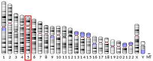
[編集]F2RL1遺伝子には2つのエクソンが含まれ、ヒトの組織で広く発現している。ヒトのPAR2のアミノ酸配列はマウスの配列と83%同一である[12]。
活性化機構
[編集]
PAR2は、Gタンパク質共役受容体ファミリーのメンバーであり、プロテアーゼ活性化受容体(PAR)ファミリーのメンバーでもある。PAR2はいくつかの異なる内在性・外因性プロテアーゼによる切断によって活性化される。PAR2は、細胞外のN末端領域に位置するアルギニンとセリンの間で切断されることで活性化される[13]。切断によって新たに露出したN末端は、活性化テザードリガンド(係留リガンド)として機能し、細胞外ループ2(ECL2)内の保存された領域に結合して受容体を活性化する[5]。受容体は、テザードリガンドの末端のアミノ酸を模倣したペプチド配列によって、タンパク質分解とは無関係に活性化される[14]。また、シグナル伝達と関係していないプロテアーゼによる他の部位での切断によって、受容体はプロテアーゼへの曝露に応答しなくなる[5]。トリプシンはPAR2を切断して炎症性シグナル伝達を開始する主要な酵素である。トロンビンも高濃度ではPAR2を切断することが示されている[15]。PAR2を切断する他の酵素としてはマスト細胞の主要なプロテアーゼであるトリプターゼがあり、PAR2のタンパク質分解によってカルシウムシグナルの伝達と増殖を誘導する[16]。PARはカリクレインの基質としても同定されており、カリクレインはさまざまな炎症過程や腫瘍形成過程に関係している。PAR2の場合、カリクレイン-4、-5、-6、-14が特に重要である[17]。疾患条件下では、PAR2によってTLR4[18]やEGFR[19]がトランス活性化されることが知られている。
機能
[編集]さまざまな細胞や組織でPARの機能の解明するために多くの研究が行われている[20]。ヒトの気道と肺実質では、PAR2は線維芽細胞の増殖の増大[21]とIL-6、IL-8、PGE2、カルシウムレベルの上昇[22]を担う。マウスでは、血管拡張に関与する[23]。PAR1ともに、PAR2の調節異常はがん細胞の浸潤性に関与している[24]。
アゴニストとアンタゴニスト
[編集]PAR2の強力かつ選択的な低分子アゴニストとアンタゴニストが発見されている[25][26][27]。
PAR2には機能的選択性(functional selectivity)が生じることがあり、異なるプロテアーゼが異なる部位でPAR2を切断することでbiased signaling(バイアスのあるシグナル伝達)が生じる[28]。合成低分子リガンドもbiased signalingを調節し、異なる機能的応答をもたらす[29]。
これまでに、PAR2は2つの異なるアンタゴニストとの共結晶構造が得られており[30]、変異導入や構造ベースのドラッグデザインによるアゴニスト(内在性リガンドであるSLIGKV)結合状態のモデリングが行われている[31]。
出典
[編集]- ^ a b c GRCh38: Ensembl release 89: ENSG00000164251 - Ensembl, May 2017
- ^ a b c GRCm38: Ensembl release 89: ENSMUSG00000021678 - Ensembl, May 2017
- ^ Human PubMed Reference:
- ^ Mouse PubMed Reference:
- ^ a b c Heuberger, Dorothea M.; Schuepbach, Reto A. (2019). “Protease-activated receptors (PARs): mechanisms of action and potential therapeutic modulators in PAR-driven inflammatory diseases”. Thrombosis Journal 17: 4. doi:10.1186/s12959-019-0194-8. ISSN 1477-9560. PMC 6440139. PMID 30976204.
- ^ “Diet-induced obesity, adipose inflammation, and metabolic dysfunction correlating with PAR2 expression are attenuated by PAR2 antagonism”. FASEB Journal 27 (12): 4757–67. (December 2013). doi:10.1096/fj.13-232702. PMID 23964081.
- ^ “Tissue factor-protease-activated receptor 2 signaling promotes diet-induced obesity and adipose inflammation”. Nature Medicine 17 (11): 1490–7. (October 2011). doi:10.1038/nm.2461. PMC 3210891. PMID 22019885.
- ^ “Protease-Activated Receptors in the Intestine: Focus on Inflammation and Cancer” (英語). Frontiers in Endocrinology 10: 717. (2019). doi:10.3389/fendo.2019.00717. PMC 6821688. PMID 31708870.
- ^ “PAR2 induces ovarian cancer cell motility by merging three signalling pathways to transactivate EGFR”. British Journal of Pharmacology 178 (4): 913–932. (November 2020). doi:10.1111/bph.15332. PMID 33226635.
- ^ “Protease and protease-activated receptor-2 signaling in the pathogenesis of atopic dermatitis”. Yonsei Medical Journal 51 (6): 808–22. (November 2010). doi:10.3349/ymj.2010.51.6.808. PMC 2995962. PMID 20879045.
- ^ “Proteinase-activated receptor-2 in the skin: receptor expression, activation and function during health and disease”. Drug News & Perspectives 21 (7): 369–81. (September 2008). doi:10.1358/dnp.2008.21.7.1255294. PMID 19259550.
- ^ “Entrez Gene: F2RL1 coagulation factor II (thrombin) receptor-like 1”. 2021年4月29日閲覧。
- ^ “Protease-activated receptors and their biological role - focused on skin inflammation”. The Journal of Pharmacy and Pharmacology 67 (12): 1623–33. (December 2015). doi:10.1111/jphp.12447. PMID 26709036.
- ^ “Potent and metabolically stable agonists for protease-activated receptor-2: evaluation of activity in multiple assay systems in vitro and in vivo”. The Journal of Pharmacology and Experimental Therapeutics 309 (3): 1098–107. (June 2004). doi:10.1124/jpet.103.061010. PMID 14976227.
- ^ “Thrombin-Mediated Direct Activation of Proteinase-Activated Receptor-2: Another Target for Thrombin Signaling”. Molecular Pharmacology 89 (5): 606–14. (May 2016). doi:10.1124/mol.115.102723. PMID 26957205.
- ^ “Mast cell tryptase stimulates human lung fibroblast proliferation via protease-activated receptor-2”. American Journal of Physiology. Lung Cellular and Molecular Physiology 278 (1): L193-201. (January 2000). doi:10.1152/ajplung.2000.278.1.l193. PMID 10645907.
- ^ “Kallikrein protease activated receptor (PAR) axis: an attractive target for drug development”. Journal of Medicinal Chemistry 55 (15): 6669–86. (August 2012). doi:10.1021/jm300407t. PMID 22607152.
- ^ “Macrophage TLR4 and PAR2 Signaling: Role in Regulating Vascular Inflammatory Injury and Repair” (英語). Frontiers in Immunology 11: 2091. (2020). doi:10.3389/fimmu.2020.02091. PMC 7530636. PMID 33072072.
- ^ “PAR2 induces ovarian cancer cell motility by merging three signalling pathways to transactivate EGFR”. British Journal of Pharmacology 178 (4): 913–932. (November 2020). doi:10.1111/bph.15332. PMID 33226635.
- ^ “Protease-activated receptors and their biological role - focused on skin inflammation”. The Journal of Pharmacy and Pharmacology 67 (12): 1623–33. (December 2015). doi:10.1111/jphp.12447. PMID 26709036.
- ^ “Mast cell tryptase stimulates human lung fibroblast proliferation via protease-activated receptor-2”. American Journal of Physiology. Lung Cellular and Molecular Physiology 278 (1): L193-201. (January 2000). doi:10.1152/ajplung.2000.278.1.l193. PMID 10645907.
- ^ “Activation of protease-activated receptor (PAR)-1, PAR-2, and PAR-4 stimulates IL-6, IL-8, and prostaglandin E2 release from human respiratory epithelial cells”. Journal of Immunology 168 (7): 3577–85. (April 2002). doi:10.4049/jimmunol.168.7.3577. PMID 11907122.
- ^ “Attenuated vasodilator effectiveness of protease-activated receptor 2 agonist in heterozygous par2 knockout mice”. PLOS ONE 8 (2): e55965. (2013-02-07). Bibcode: 2013PLoSO...855965H. doi:10.1371/journal.pone.0055965. PMC 3567012. PMID 23409098.
- ^ “Protease-activated receptors (PARs) in cancer: Novel biased signaling and targets for therapy”. Methods in Cell Biology 132: 341–58. (2016). doi:10.1016/bs.mcb.2015.11.006. PMID 26928551.
- ^ “Identification and characterization of novel small-molecule protease-activated receptor 2 agonists”. The Journal of Pharmacology and Experimental Therapeutics 327 (3): 799–808. (December 2008). doi:10.1124/jpet.108.142570. PMID 18768780.
- ^ “Novel agonists and antagonists for human protease activated receptor 2”. Journal of Medicinal Chemistry 53 (20): 7428–40. (October 2010). doi:10.1021/jm100984y. PMID 20873792.
- ^ “PAR2 Modulators Derived from GB88”. ACS Medicinal Chemistry Letters 7 (12): 1179–1184. (December 2016). doi:10.1021/acsmedchemlett.6b00306. PMC 5150695. PMID 27994760.
- ^ “Biased signaling of protease-activated receptors”. Frontiers in Endocrinology 5: 67. (2014). doi:10.3389/fendo.2014.00067. PMC 4026716. PMID 24860547.
- ^ “Biased Signaling by Agonists of Protease Activated Receptor 2”. ACS Chemical Biology 12 (5): 1217–1226. (May 2017). doi:10.1021/acschembio.6b01088. PMID 28169521.
- ^ “Structural insight into allosteric modulation of protease-activated receptor 2”. Nature 545 (7652): 112–115. (May 2017). Bibcode: 2017Natur.545..112C. doi:10.1038/nature22309. PMID 28445455.
- ^ “Structural Characterization of Agonist Binding to Protease-Activated Receptor 2 through Mutagenesis and Computational Modeling”. ACS Pharmacology & Translational Science 1 (2): 119–133. (November 2018). doi:10.1021/acsptsci.8b00019. PMC 7088944. PMID 32219208.
関連文献
[編集]- “Ion transport induced by proteinase-activated receptors (PAR2) in colon and airways”. Cell Biochemistry and Biophysics 36 (2–3): 209–14. (2003). doi:10.1385/CBB:36:2-3:209. PMID 12139406.
- “PAR-2: structure, function and relevance to human diseases of the gastric mucosa”. Expert Reviews in Molecular Medicine 4 (16): 1–17. (July 2002). doi:10.1017/S1462399402004799. PMID 14585156.
- “The emergence of proteinase-activated receptor-2 as a novel target for the treatment of inflammation-related CNS disorders”. The Journal of Physiology 581 (Pt 1): 7–16. (May 2007). doi:10.1113/jphysiol.2007.129577. PMC 2075212. PMID 17347265.
- “Molecular cloning and functional expression of the gene encoding the human proteinase-activated receptor 2”. European Journal of Biochemistry 232 (1): 84–9. (August 1995). doi:10.1111/j.1432-1033.1995.tb20784.x. PMID 7556175.
- “Evidence for the presence of a protease-activated receptor distinct from the thrombin receptor in human keratinocytes”. Proceedings of the National Academy of Sciences of the United States of America 92 (20): 9151–5. (September 1995). Bibcode: 1995PNAS...92.9151S. doi:10.1073/pnas.92.20.9151. PMC 40942. PMID 7568091.
- “Molecular cloning of a potential proteinase activated receptor”. Proceedings of the National Academy of Sciences of the United States of America 91 (20): 9208–12. (September 1994). Bibcode: 1994PNAS...91.9208N. doi:10.1073/pnas.91.20.9208. PMC 44781. PMID 7937743.
- “The proteinase activated receptor-2 (PAR-2) mediates mitogenic responses in human vascular endothelial cells”. The Journal of Clinical Investigation 97 (7): 1705–14. (April 1996). doi:10.1172/JCI118597. PMC 507235. PMID 8601636.
- “Molecular cloning, expression and potential functions of the human proteinase-activated receptor-2”. The Biochemical Journal 314 ( Pt 3) (3): 1009–16. (March 1996). doi:10.1042/bj3141009. PMC 1217107. PMID 8615752.
- “Mechanisms of desensitization and resensitization of proteinase-activated receptor-2”. The Journal of Biological Chemistry 271 (36): 22003–16. (September 1996). doi:10.1074/jbc.271.36.22003. PMID 8703006.
- “Conserved structure and adjacent location of the thrombin receptor and protease-activated receptor 2 genes define a protease-activated receptor gene cluster”. Molecular Medicine 2 (3): 349–57. (May 1996). doi:10.1007/BF03401632. PMC 2230143. PMID 8784787.
- “Interactions of mast cell tryptase with thrombin receptors and PAR-2”. The Journal of Biological Chemistry 272 (7): 4043–9. (February 1997). doi:10.1074/jbc.272.7.4043. PMID 9020112.
- “Proteinase-activated receptor-2: expression by human neutrophils”. Journal of Cell Science 110 ( Pt 7) (7): 881–7. (April 1997). PMID 9133675.
- “Characterization of protease-activated receptor-2 immunoreactivity in normal human tissues”. The Journal of Histochemistry and Cytochemistry 46 (2): 157–64. (February 1998). doi:10.1177/002215549804600204. PMID 9446822.
- “Protease-activated receptor genes are clustered on 5q13”. Blood 92 (1): 25–31. (July 1998). doi:10.1182/blood.V92.1.25.413k41_25_31. PMID 9639495.
- “Proteinase-activated receptor-2 in human skin: tissue distribution and activation of keratinocytes by mast cell tryptase”. Experimental Dermatology 8 (4): 282–94. (August 1999). doi:10.1111/j.1600-0625.1999.tb00383.x. PMID 10439226.
- “Cellular localization of membrane-type serine protease 1 and identification of protease-activated receptor-2 and single-chain urokinase-type plasminogen activator as substrates”. The Journal of Biological Chemistry 275 (34): 26333–42. (August 2000). doi:10.1074/jbc.M002941200. PMID 10831593.
- “Proteolysis of the exodomain of recombinant protease-activated receptors: prediction of receptor activation or inactivation by MALDI mass spectrometry”. Biochemistry 39 (35): 10812–22. (September 2000). doi:10.1021/bi0003341. PMID 10978167.
- “Protease-activated receptors in human airways: upregulation of PAR-2 in respiratory epithelium from patients with asthma”. The Journal of Allergy and Clinical Immunology 108 (5): 797–803. (November 2001). doi:10.1067/mai.2001.119025. PMID 11692107.
- “Trypsin induces activation and inflammatory mediator release from human eosinophils through protease-activated receptor-2”. Journal of Immunology 167 (11): 6615–22. (December 2001). doi:10.4049/jimmunol.167.11.6615. PMID 11714832.
- “Activation of protease-activated receptor (PAR)-1, PAR-2, and PAR-4 stimulates IL-6, IL-8, and prostaglandin E2 release from human respiratory epithelial cells”. Journal of Immunology 168 (7): 3577–85. (April 2002). doi:10.4049/jimmunol.168.7.3577. PMID 11907122.
- “Protease-activated receptor 2 has pivotal roles in cellular mechanisms involved in experimental periodontitis”. Infection and Immunity 78 (2): 629–38. (February 2010). doi:10.1128/IAI.01019-09. PMC 2812191. PMID 19933835.
関連項目
[編集]外部リンク
[編集]- “Protease-Activated Receptors: PAR2”. IUPHAR Database of Receptors and Ion Channels. International Union of Basic and Clinical Pharmacology. 2021年4月29日閲覧。


